
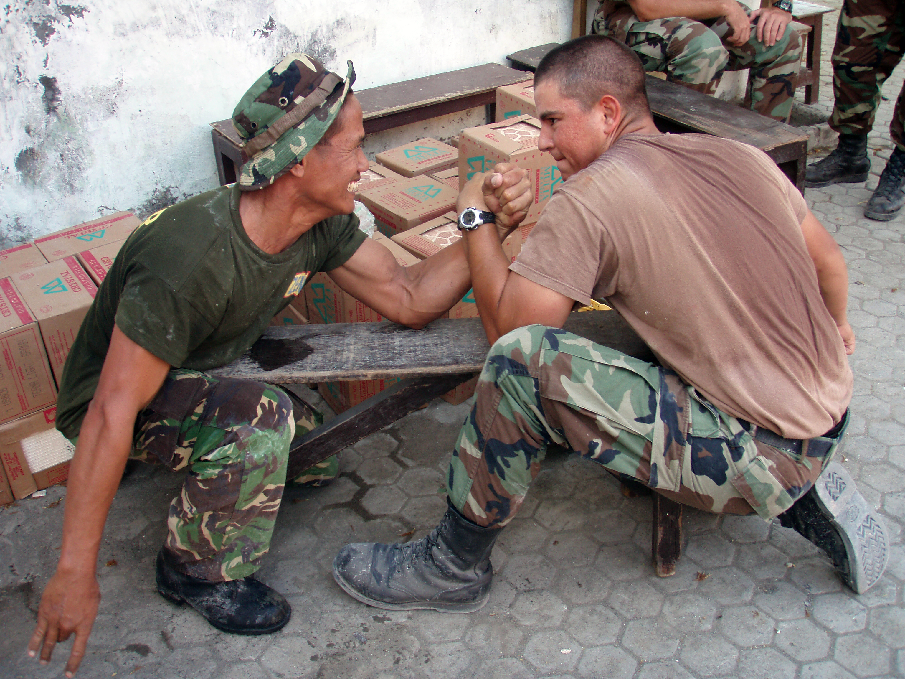
A sport like arm-wrestling depends on muscle contractions. Arm wrestlers must contract muscles in their hands and arms and keep them contracted to resist their opponent's opposing force. The wrestler whose muscles can contract with greater force wins the match.
Excluding reflexes, all skeletal muscle contractions occur as a result of conscious effort originating in the brain. The brain sends electrochemical signals through the somatic nervous system to motor neurons that innervate muscle fibers (to review how the brain and neurons function, see the chapter Nervous System). A single motor neuron with multiple axon terminals can innervate multiple muscle fibers, thereby causing them to contract at the same time. The connection between a motor neuron axon terminal and a muscle fiber occurs at a neuromuscular junction site. This is a chemical synapse where a motor neuron transmits a signal to muscle fiber to initiate a muscle contraction. The process by which a signal is transmitted at a neuromuscular junction is illustrated in Figure \(\PageIndex\). The sequence of events begins when an action potential is initiated in the cell body of a motor neuron, and the action potential is propagated along the neuron’s axon to the neuromuscular junction. Once the action potential reaches the end of the axon terminal, it causes the neurotransmitter acetylcholine (ACh) from synaptic vesicles in the axon terminal. The ACh molecules diffuse across the synaptic cleft and bind to the muscle fiber receptors, thereby initiating a muscle contraction. Muscle contraction is initiated with the depolarization of the sarcolemma caused by the sodium ions' entrance through the sodium channels associated with the ACh receptors. 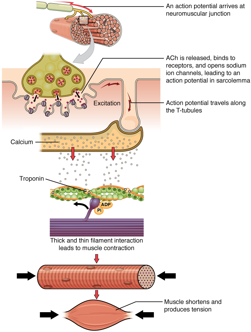 Things happen very quickly in the world of excitable membranes (think about how quickly you can snap your fingers as soon as you decide to do it). Immediately following depolarization of the membrane, it repolarizes, re-establishing the negative membrane potential. Meanwhile, the ACh in the synaptic cleft is degraded by the enzyme acetylcholinesterase (AChE). The ACh cannot rebind to a receptor and reopen its channel, which would cause unwanted extended muscle excitation and contraction. Propagation of an action potential along the sarcolemma enters the T-tubules. For the action potential to reach the membrane of the Sarcoplasmic Reticulum (SR), there are periodic invaginations in the sarcolemma, called T-tubules (“T” stands for “transverse”). The arrangement of a T-tubule with the membranes of SR on either side is called a triad (Figure \(\PageIndex\)). The triad surrounds the cylindrical structure called a myofibril, which contains actin and myosin. The T-tubules carry the action potential into the interior of the cell, which triggers the opening of calcium channels in the membrane of the adjacent SR, causing \(\text^<++>\) to diffuse out of the SR and into the sarcoplasm. It is the arrival of \(\text^<++>\) in the sarcoplasm that initiates contraction of the muscle fiber by its contractile units, or sarcomeres.
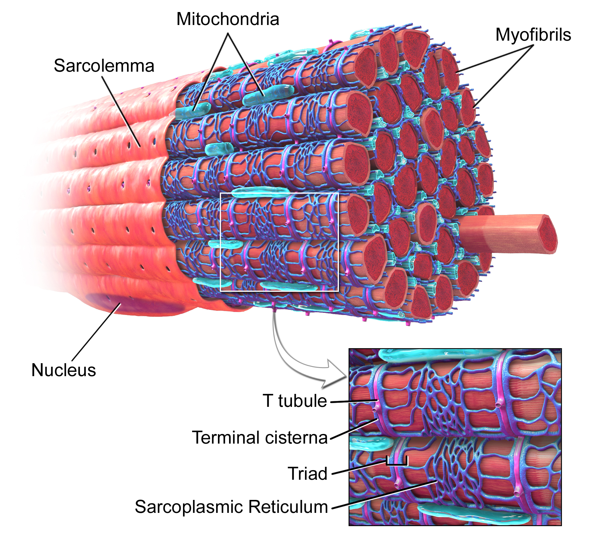
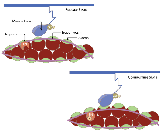
Although the term excitation-contraction coupling confuses or scares some students, it comes down to this: for a skeletal muscle fiber to contract, its membrane must first be “excited”—in other words, it must be stimulated to fire an action potential. The muscle fiber action potential, which sweeps along the sarcolemma as a wave, is “coupled” to the actual contraction through the release of calcium ions (\(\text^<++>\)) from the SR. Once released, the \(\text^<++>\) interacts with the shielding proteins, troponin and tropomyosin complex, forcing them to move aside so that the actin-binding sites are available for attachment by myosin heads. The myosin then pulls the actin filaments toward the center, shortening the muscle fiber. In skeletal muscle, this sequence begins with signals from the somatic motor division of the nervous system. In other words, the “excitation” step in skeletal muscles is always triggered by signaling from the nervous system.
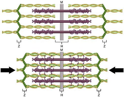
Once the muscle fiber is stimulated by the motor neuron, actin, and myosin protein filaments within the skeletal muscle fiber slide past each other to produce a contraction. The sliding filament theory is the most widely accepted explanation for how this occurs. According to this theory, muscle contraction is a cycle of molecular events in which thick myosin filaments repeatedly attach to and pull on thin actin filaments, so they slide over one another. The actin filaments are attached to Z discs, each of which marks the end of a sarcomere. The sliding of the filaments pulls the Z discs of a sarcomere closer together, thus shortening the sarcomere. As this occurs, the muscle contracts.
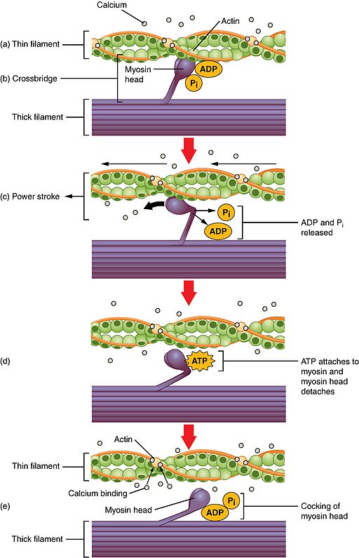
Crossbridge cycling is a sequence of molecular events that underlies the sliding filament theory. There are many projections from the thick myosin filaments, each of which consists of two myosin heads (you can see the projections and heads in Figures \(\PageIndex\) and \(\PageIndex\)). Each myosin head has binding sites for ATP (or ATP hydrolysis products: ADP and Pi) and actin. The thin actin filaments also have binding sites for the myosin heads—a cross-bridge forms when a myosin head binds with an actin filament. The process of cross-bridge cycling is shown in Figure \(\PageIndex\). A cross-bridge cycle begins when the myosin head binds to an actin filament. ADP and Pi are also bound to the myosin head at this stage. Next, a power stroke moves the actin filament inward toward the sarcomere center, thereby shortening the sarcomere. At the end of the power stroke, ADP and Pi are released from the myosin head, leaving the myosin head attached to the thin filament until another ATP binds to the myosin head. When ATP binds to the myosin head, it causes the myosin head to detach from the actin filament. ATP is again split into ADP and Pi and the energy released is used to move the myosin head into a "cocked" position. Once in this position, the myosin head can bind to the actin filament again, and another cross-bridge cycle begins.
This page titled 15.4: Muscle Contraction is shared under a CK-12 license and was authored, remixed, and/or curated by Suzanne Wakim & Mandeep Grewal via source content that was edited to the style and standards of the LibreTexts platform.
LICENSED UNDER